 Basic Bone Analysis Application
Overview
MicroView's Basic Bone Analysis Application performs a variety of analysis upon a selected region of interest within an image. The application is designed specifically for analysis of CT images of bone. The choice of which functions to perform on a given ROI, can be selected prior to any calculation. The results could be stored or appended in user specified plain text files.
The Basic Bone Analysis Application contains the following analysis tools:
- BMD, which reports the bone mineral density (BMD), bone volume fraction (BVF), bone mineral content, and various other statistics;
- Stereology, which reports Euler index, bone volume fraction, bone surface to bone volume ratio, trabecular plate thickness, trabecular plate number, trabecular plate separation and various other measures. The Euler index is a measure of the connectivity of a trabecular structure.
Tip
The commercial package Advanced Bone Analysis Application contains analysis tools for SMI, Anisotropy, and Stereology. It also offers project and study management tools and support PDF and Excel outputs.
Using the Basic Bone Analysis Application
- Activate the Basic Bone Analysis Application by selecting
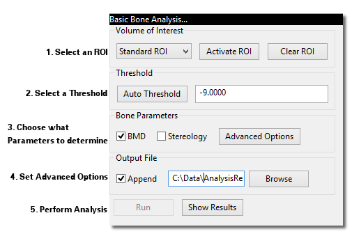
Analyze→Bone Analysis...from MicroView's menu, or by clicking on the Bone Analysis button in the Applications group of MicroView's toolbar. - If an ROI has not already been selected, first activate an ROI plugin and select a ROI. As a convenience, the list of available ROI plugins is displayed in the choice-box in the upper left of the plugin: Choose an ROI tool in the available drop-down list, then hit the Activate button to launch the tool.
- Once a ROI has been selected, enter a gray-level threshold value, that will be used to discriminate bone from none-bone voxels in the image. Either type in a threshold value in the entry field in the Threshold section or click the Auto Threshold button, to determine a best-guess threshold value automatically. If you would like to verify that the automatically selected value is appropriate, generate a histogram of the image contained within the ROI, then hit the Auto Threshold button on the histogram plot window.
- In the Bone Parameters section select the type of analysis desired. Click the Advanced Options... button to modify the default settings. The Advanced options will be discussed in more detail later in this section.
- In the Output File section select the name of the file where the results will be stored. The results are written to a plain text file. These results are presented for review after each successful execution. It is also possible to append the results to an existing text file by checking the Append checkbox button.
- Click the Run button to perform the analysis.
Advanced Options
The Advanced Options allows the user to modify the default settings. To display the Advanced Options dialog click the Advanced Options... button. In the Advanced Options dialog there are several tabs:
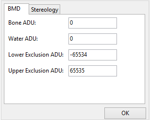
- The BMD tab has several variables that can be edited manually. By default the values for Bone ADU (arbitrary density unit) and Water ADU are set to the calibration constants found in the header of the currently loaded image file (not all file types support this). The Lower Exclusion ADU is a gray scale value below which voxels are not included in the bone equivalent mass calculation. Similarly the Upper Exclusion ADU is a Gray scale value above which voxels are not included in the bone equivalent mass calculation. The upper and lower exclusion ADU should be set to exclude air and metal, respectively, in the bone mass calculation.
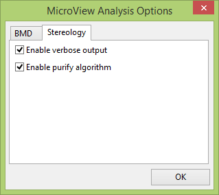
- The Stereology tab, has an option to enable verbose output to display additional measures, and an option to enable the purify algorithm. To obtain meaningful results from Stereology, the image should be passed through this purification filter first. The purify algorithm removes spurious unconnected region.
- After all the modifications to the Advanced Options have been made, click the OK button to close the dialog.
Specific Tool Information
Bone Composition Measurement Tool
Bone Composition
This tool performs a virtual biopsy and "ashing" to determine bone mineral content non-destructively. Image data derived from the Locus family of CT scanners may be calibrated to standard CT number, measured in Hounsfield Units (HU), and furthermore calibrated to permit determination of equivalent mass of hydroxyappetite. Results are reported as bone mineral fraction (BVF) or bone mineral density (BMD) in units of mg (HyAp)/cm3. To use this tool, launch the Advanced Bone Analysis application, define a 3D ROI, then select a threshold that discriminates bone from soft tissue. Prior to hitting the Run button, if required, click the Advanced Options... button to modify the BMD tool settings:
- Enter a value for
Bone ADU - Enter a value for
Water ADU - Enter a value for
Lower Exclusion ADU - Enter a value for
Upper Exclusion ADU
Stereology Tool
Stereology
MicroView can perform a simple stereology analysis of a 3D bone image. The stereology tool measures trabecular structure using similar techniques to those implemented in classical histomorphometry. 2D techniques determine estimates of trabecular thickness, spacing and density. At the same time, trabecular connectivity is quantified by calculating the Euler number for the trabecular structure. Finally, the bone surface area to volume ratio is also calculated.
To perform a stereology analysis on a CT image of a bone, launch the Advanced Bone Analysis application, define a 3D ROI, then select a threshold that discriminates bone from soft tissue. Prior to hitting the Run, if required, click the Advanced button to modify the Stereology tool settings:
- Optionally click on the
Enable Purifyoption to pass the selected image through a purification algorithm, first, before performing the stereology analysis. The purify algorithm removes isolated bony spicules and fills encapsulated marrow spaces. Note: When the purify algorithm is enabled, select a ROI where at least one of the voxels at the boundary of the ROI is equal to or above the threshold (ie. the trabecular bone must intersect the boundaries of the ROI). Passing a clipped image, through the purify filter, which has no boundary voxels equal to or above the threshold will produce a blank image by the filter. - Optionally uncheck the Enable verbose output to disable the verbose output.